Systematic palaeontology
Saurischia Seeley, 188737
Theropoda Marsh, 188138
Tetanurae Gauthier, 198639
Megaraptora Benson, Carrano, and Brusatte, 201033
Megaraptoridae Novas, Agnolín, Ezcurra, Porfiri, and Canale, 201334
Joaquinraptor casali gen. et sp. nov.
Etymology
Joaquín, in tribute to the son of the first author (L.M.I.) and the informal name given to the locality when the skeleton of the taxon was discovered (Valle Joaquín); Latin, raptor, thief. Specific epithet casali in recognition of Dr. Gabriel Andrés Casal for his many contributions to knowledge of the Cretaceous palaeontology and geology of central Patagonia (including the formal recognition and naming of the formation from which this megaraptorid was recovered).
Holotype
UNPSJB-PV 1112, a partially articulated partial skeleton of a single individual that includes the mostly disarticulated partial skull (right maxilla, skull roof and braincase, probable right postorbital, right quadrate, both dentaries, in situ and isolated teeth), complete or nearly complete elements of the postcranial axial (atlantal intercentrum, three caudal vertebrae, dorsal ribs, other rib and probable gastralium fragments, haemal arch) and appendicular (left scapulocoracoid, humerus, radius, and ulna, right manual ungual II, left femur, much of the right tibia, the distal end of the probable right pedal phalanx III-3, the right pedal ungual III, and the possible partial left pedal phalanx IV-1) skeletons, and numerous indeterminate fragments.
Locality and horizon
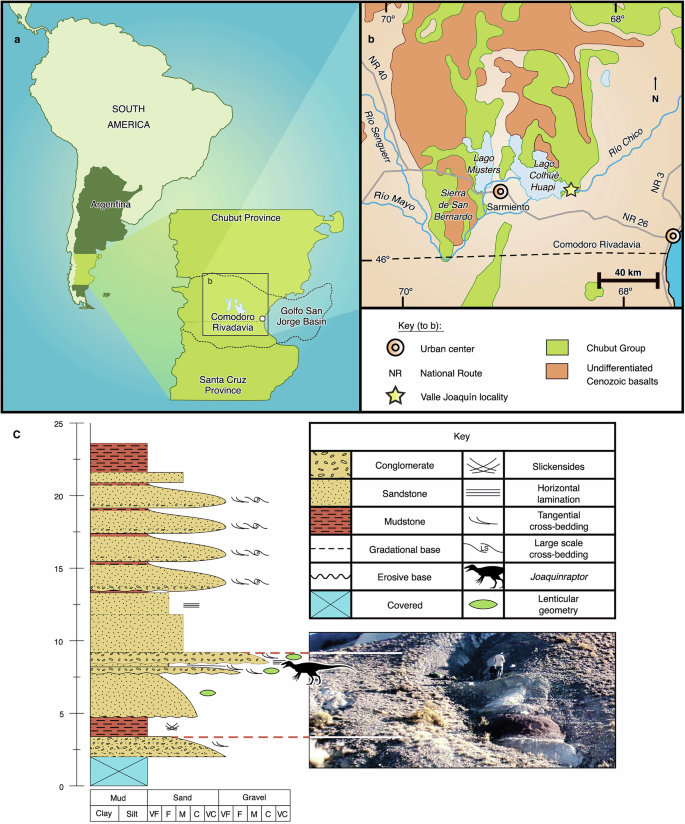
Valle Joaquín, headwaters of the Río Chico, east of the southeastern shore of Lago Colhué Huapi, Chubut Province, central Patagonia, Argentina. Upper (Maastrichtian, probably upper Maastrichtian) stratum of the Upper Cretaceous Lago Colhué Huapi Formation (see below; also Fig. 1 and Supplementary Figs. 1 and 2).
a Location of the study area in south-central Chubut Province, central Patagonia, Argentina. b Simplified geologic map showing the informally named Valle Joaquín locality in the Upper Cretaceous Lago Colhué Huapi Formation (part of the Chubut Group) that yielded Joaquinraptor casali gen. et sp. nov. c Stratigraphic column and photograph of Valle Joaquín showing the position of the horizon of the Lago Colhué Huapi Formation that yielded Joaquinraptor. Abbreviations: C coarse, F fine, M medium, VC very coarse, VF very fine.
Diagnosis
The holotype of Joaquinraptor casali is considered to represent a sexually but possibly not somatically mature individual based on the fusion of braincase sutures and the results of our osteohistological analysis, which reveal a reduction in the distance between growth marks in the outer bony cortex of limb bones (see below). Joaquinraptor is regarded as a member of Megaraptoridae based on its possession of the following suite of synapomorphies of the clade: (1) reduced anteriormost dentary alveolus35; (2) absence of mesial denticles from tooth crowns34,35,40,41; (3) olecranon process of ulna mediolaterally compressed, blade-like, and subtriangular in lateral view; and (4) ulnar shaft with well-developed, proximodistally oriented lateral crest or tuberosity21,34,35,36. Joaquinraptor is diagnosed by the following autapomorphies (see also Supplementary Note 4 and Supplementary Figs. 3–7): (1) parietals strongly compressed transversely, narrower than frontals; (2) anterior margin of supratemporal fossa with sigmoid contour; (3) braincase lacking median septum in pneumatic recess (probably, but see below); (4) rugose atlantal intercentrum with well-marked lips and rectangular ventral contour; (5) middle or posterior caudal vertebral centra with well-marked elliptical lateral fossae that occupy more than half the lateral surfaces; (6) humerus with subrectangular deltopectoral crest; ulna with (7) proximodistally enlarged olecranon process, (8) vertical ridge on medial surface of proximal end, (9) nearly straight shaft (but see below), and (10) medially oriented, anteroposteriorly compressed distal end; and (11) distal end of radius crescent-shaped. Differs from the only other named Maastrichtian (probably early Maastrichtian and therefore stratigraphically older; see refs. 42,43,44) megaraptorid species Maip macrothorax in having a coracoid with a subglenoid ridge, posteroventral fossa, and hooked posteroventral process. The preserved dorsal ribs of Joaquinraptor and Maip also exhibit morphological differences, though these could potentially be due to non-equivalence in serial position rather than taxonomic distinction.
This published work and the nomenclatural acts it contains have been registered in ZooBank, the proposed online registration system for the International Code of Zoological Nomenclature (ICZN). The ZooBank LSIDs (Life Science Identifiers) can be resolved and the associated information viewed through any standard web browser by appending the LSID to the prefix “http://zoobank.org/”. The LSIDs for this publication are: urn:lsid:zoobank.org:pub:468D27F1-2076-4187-911C-E70028218787; urn:lsid:zoobank.org:act:AED2BB3B-C081-445C-A081-C357E7CEC580; urn:lsid:zoobank.org:act:3AA601F2-5DFC-452B-9609-E83C913F8C2D.
Age
Geological studies of the uppermost part of the Lago Colhué Huapi Formation, particularly those of palynomorphs, support a late Maastrichtian age for these strata, probably close to the Cretaceous/Palaeogene boundary45,46. The palaeoclimate of central Patagonia during deposition of this part of the formation is suggested to be warm with at least seasonal rainfall45. On the other hand, palaeoclimatic studies of the middle (Santonian –?lower Maastrichtian) section of the Lago Colhué Huapi Formation indicate a seasonally dry or semiarid climate47. Recent analysis of clays from the Joaquinraptor quarry indicates a composition similar to that of the top of the formation (see Supplementary Note 3). Therefore, this megaraptorid was found at a stratigraphic level that is more related to those of the top of this unit than those of the middle section, supporting a Maastrichtian (and probably late Maastrichtian) age for this specimen.
The oldest megaraptorans are Phuwiangvenator yaemniyomi and Vayuraptor nongbualamphuensis from Thailand11,12,13, both probably late Valanginian–early Hauterivian in age48. The youngest records of the clade are from the Campanian–Maastrichtian of Argentina, Chile, and possibly Brazil8,22,23,49,50,51,52,53. Nevertheless, these latter megaraptorans probably date to the Campanian–early Maastrichtian interval8,42,43,44,52,53,54 and are thus similar in age to the fragmentary megaraptorid fossils (mostly manual unguals) recovered from the middle section of the Lago Colhué Huapi Formation7,31,55. Therefore, Joaquinraptor seemingly constitutes the stratigraphically youngest representative of Megaraptora yet discovered anywhere in the world. Interestingly, what are probably both the oldest7 and youngest (Joaquinraptor) definitive South American megaraptorids known at present come from strata of the Golfo San Jorge Basin of central Patagonia.
Osteological description
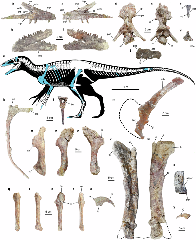
The megaraptoran fossil record is generally depauperate; therefore, not many specimens within the group preserve overlapping skeletal elements. In this context, Joaquinraptor casali is important because it is represented by a well-preserved partial skeleton that includes cranial, postcranial axial, and appendicular elements (Fig. 2a and Tables 1, 2), allowing relatively extensive comparisons with other megaraptoran taxa (see Supplementary Notes 5 and 6 and Supplementary Figs. 8–18). This is the case for the subtriangular maxilla (Fig. 2b, c), which is otherwise preserved only in a juvenile specimen of Megaraptor namunhuaiquii (MUCPv 595; see ref. 6). In Joaquinraptor, the anterior part of the antorbital fossa is perforated by a poorly delimited, fossa-like structure that may represent the confluent maxillary fenestra and promaxillary foramen. The morphology and configuration of these probable pneumatic excavations closely resembles that in Megaraptor, supporting this condition as a synapomorphy of Megaraptora or one of its subclades instead of an autapomorphy of the latter genus (contra6). Similarly, in both these megaraptorids, the medial maxillary shelf bifurcates posteriorly, forming a deep, “horizontal V”-shaped, well-defined antrum (=posterior anteromaxillary fenestra; see ref. 56), which may represent another megaraptoran synapomorphy. The maxilla of Joaquinraptor differs from that of Megaraptor in having a substantially taller and blunter anterior process (=anterior ramus), an anteroposteriorly narrower anterior part of the antorbital fossa, an acute anterior margin of the antorbital fenestra, and a dorsoventrally thicker ascending process, although whether these distinctions are of taxonomic significance rather than reflective of the differing ontogenetic stages of UNPSJB-PV 1112 and MUCPv 595 is unknown at present. Interestingly, in being low, elongate, and subtriangular, the maxilla of Joaquinraptor is also somewhat similar to that of the penecontemporaneous large-bodied unenlagiine theropod Austroraptor cabazai from northern Patagonia57. Nevertheless, the maxilla of the new megaraptorid differs from that of Austroraptor in, among other features, the degree of elongation of the antorbital region (which is pronounced in the unenlagiine), the shape of the antorbital fossa, and the number of alveoli (which is substantially higher in the unenlagiine). Although the surfaces of the maxillary interdental plates are slightly eroded in the Joaquinraptor holotype, they appear to be unfused, unlike those of Fukuiraptor kitadaniensis2. The interdental plates are triangular in shape and subequal in size, as in the juvenile Megaraptor (MUCPv 595). On the other hand, allosauroids more derived than Sinraptor dongi have larger interdental plates6.
a Skeletal reconstruction of Joaquinraptor in left lateral view with preserved elements in blue (some reversed from right side) (modified and updated from Lamanna et al.7 [these authors’ Fig. 1e], which was in turn modified by A. McAfee from an original illustration by T.K. Robinson). Right maxilla in lateral (b) and medial (c) views. Skull roof, braincase, and atlantal intercentrum in dorsal (d) and ventral (e) views. f Probable right postorbital in lateral view. g Right quadrate in anterior view. Right and left dentaries in lateral and medial views (right dentary in lateral view and left dentary in medial view in h; opposite in i). j Two articulated middle or posterior caudal vertebrae in right lateral view. k Dorsal rib in anterior view. l Anterior haemal arch in anterior view. m Left scapulocoracoid in lateral view. Left humerus in anterolateral (n), lateral (o), and medial (p) views. Left radius in anterior (q) and lateral (r) views. Left ulna in lateral (s) and posterior (t) views. u Right manual ungual II (=manual phalanx II-3) in lateral view. Left femur in anterior (v) and posterior (w) views. x Distal right tibia in anterior view. y Right pedal ungual III (=pedal phalanx III-4) in medial view. Dashed lines indicate missing areas of scapulocoracoid and femur. Abbreviations: anfe antorbital fenestra, anfo antorbital fossa, anp anterior process, aspar articular surface for ascending process of astragalus, at anterior trochanter, ati atlantal intercentrum, cap capitulum, cor coracoid, dp deltopectoral crest, et extensor tubercle, f frontal, fh femoral head, fo lateral fossa, ft flexor tubercle, ftr fourth trochanter, gl glenoid fossa, gt greater trochanter, hc haemal canal, hop horizontal process, lc lateral condyle, lms longitudinal medial shelf, lt lateral tuberosity, mg Meckelian groove, mf maxillary fenestra, mhc medial hemicondyle, mm medial malleolus, oc occipital condyle, op olecranon process, p parietal, pa posterior antrum, pap palatal process, pnr pneumatic recess, po articular surface for postorbital, pop paroccipital process, pot postorbital tuberosity, poz postzygapophysis, pp posterior process, prf promaxillary foramen, prz prezygapophysis, scb scapular blade, stf supratemporal fenestra, tub tuberculum, uc ulnar condyle, vg vascular groove, vp ventral process.
The braincase (Fig. 2d, e) is anteroposteriorly longer and more transversely compressed than that of the reportedly subadult holotype of the megaraptorid Murusraptor barrosaensis (MCF-PVPH 411; see ref. 25)—which is, at present, the only other almost complete megaraptoran braincase known—and still more narrow and elongate than the partial braincase of the aforementioned juvenile Megaraptor (MUCPv 595; see ref. 6). No sutures are visible (except for the interparietal suture, see immediately below), which accords with osteohistological evidence that indicates that the Joaquinraptor holotype was sexually and possibly somatically mature at the time of death (minimum age of 19 years; see below). Nevertheless, although it has possibly been exaggerated by potential taphonomic artifacts, a well-marked, non-interdigitated, open interparietal suture is evident, as is also the case in Murusraptor; this feature may therefore characterize Megaraptoridae27. The parietals are more transversely constricted than in Murusraptor, closely resembling the condition in tyrannosauroids36; this feature is therefore considered an autapomorphy of Joaquinraptor within Megaraptora (Supplementary Fig. 3). The frontals are subquadrangular in shape, as in other megaraptorans6,18,24,25. In lateral view, the posterior region of the ventrolaterally oriented frontals occupies a more dorsal position than the anterior portion, conferring a step-like morphology similar to that of Murusraptor (see ref. 27) and the probable megaraptorid MCF-PVPH 320 (see ref. 24); this feature may therefore represent another synapomorphy of Megaraptora or a clade therein. Anteriorly, there is a wide, anteriorly projected supratemporal fossa similar to that seen in Megaraptor and Murusraptor, although it differs from those of these other megaraptorids in having a sigmoid contour in dorsal view. Conversely, in allosauroids such as Acrocanthosaurus atokensis, Allosaurus fragilis, and Carcharodontosaurus saharicus, the supratemporal fossa is anteroposteriorly short36 and its anterior margin exhibits a more ovoid aspect. The ventrolateral inclination of the frontals from the midline of the skull roof, forming a triangular cross-section, has been regarded as an autapomorphy of Murusraptor (see ref. 27), but may instead represent a synapomorphy of Megaraptoridae as it is also present in Joaquinraptor. Ventrally, the median septum that divides the basisphenoid recess that is described in Megaraptor6 and Murusraptor27 appears to be absent in Joaquinraptor, although this could potentially be an artifact of preservation. The more complete of the two basipterygoid processes is robust and hooks posteriorly in lateral view, unlike those of Murusraptor that are shorter and mostly ventrally oriented.
The bone tentatively interpreted as the right postorbital (Fig. 2f) is almost complete, missing only parts of the anterior and posterior processes, though it has probably also experienced some plastic deformation. It is anteroposteriorly and mediolaterally narrower than the postorbitals of Aerosteon riocoloradensis4,30, Murusraptor25, and Orkoraptor burkei22. Compared with these megaraptorids, the anterior process appears shorter and the ventral process straighter (i.e., the latter is not ‘cradle-shaped’). Joaquinraptor shares with Aerosteon and Murusraptor the presence of a small tuberosity on the anterior margin of the ventral process that is well exposed in lateral and anterior views (see ref. 30, these authors’ Fig. 4a, d, “rugose bump”). Furthermore, both Joaquinraptor and Aerosteon possess a well-marked foramen on the dorsal surface of the postorbital, situated relatively close to the frontal contact.
The condylar (ventral) portion of the right quadrate is preserved (Fig. 2g). In overall morphology, it resembles the quadrate of Aerosteon more than that of Murusraptor, which is generally more gracile. There is a well-marked, enclosed posterior pneumatic foramen (sensu58) on the posterior surface, as in other megaraptorids and many other coelurosaurs58. In Joaquinraptor, however, this foramen is placed on the lateral part of the quadrate adjacent to the quadratojugal contact, whereas in Aerosteon and Murusraptor it is more centrally placed.
An isolated maxillary tooth of Joaquinraptor (Supplementary Fig. 17) is proportionally apicobasally taller than those of Murusraptor. As in other megaraptorids, the tooth is labiolingually compressed, the distal margin of the crown is strongly concave in labial and lingual views, and the distal denticles are subrectangular to rectangular. The mesial margin lacks a serrated carina, as in most other South American megaraptorids40.
The Joaquinraptor dentaries (Fig. 2h, i) are elongate, mediolaterally compressed, and shallow in lateral view, as in Australovenator wintonensis (AODF 604)59. Furthermore, both these megaraptorids and the early branching megaraptoran Fukuiraptor2 share a reduced symphyseal facet and an anterior dentary margin that trends anterodorsally–posteroventrally. However, in overall morphology, the dentary of Joaquinraptor is more robust than that of Australovenator. Slightly pronounced primary and secondary neurovascular foramina are evident on the lateral surface. White et al.59 suggested that the absence of these foramina may be autapomorphic of Australovenator, contrasting with the original description of this taxon (see ref. 5). Similar neurovascular openings were described and figured in dentary fragments of Fukuiraptor2; therefore, the presence of slightly marked neurovascular foramina that are not enclosed or delimited by grooves may represent a synapomorphy of Megaraptora or one of its subclades (e.g., Megaraptoridae). Both Joaquinraptor dentaries preserve 17 alveoli—two fewer than in Australovenator—although this could be a taphonomic artifact caused by erosion of the posteriormost portions of these bones. Unlike those of Fukuiraptor and Australovenator, the dentary interdental plates are unfused.
The atlantal intercentrum is articulated with the occipital condyle (Fig. 2e and Supplementary Fig. 3b), such that most of its anterior features cannot be described in detail; however, it is rugose and disc-shaped, differing from its more crescentic counterparts in the megaraptorans Aerosteon4,30, Orkoraptor22, and Phuwiangvenator11. Ventrally, the intercentrum exhibits well-marked rims and is rectangular in shape, unlike other megaraptorans in which this element is preserved.
The three preserved caudal vertebrae belong to the middle or posterior section of the tail, being substantially longer anteroposteriorly than tall dorsoventrally (see Table 1). The centra (Fig. 2j) are hourglass-shaped in ventral view, and their lateral surfaces are excavated by well-developed, elliptical fossae, as in many other definitive or possible megaraptorans (e.g.,4,7,22,30,49,50,60,61); however, the fossae seen in Joaquinraptor are proportionally very large, encompassing more than half the length of the lateral surfaces, a unique feature of this megaraptorid (Supplementary Fig. 4). Dorsal ribs (Fig. 2k) are pneumatized and generally resemble those of other megaraptorids, such as Australovenator5, Maip macrothorax8, and Murusraptor25. Nevertheless, and although this could potentially be due to differences in serial position, the preserved ribs of Joaquinraptor appear more gracile than those of Maip, and also do not exhibit the prominent, subtriangular medial flange present in the first dorsal rib of that southern Patagonian taxon.
The anterior haemal arch (Fig. 2l) is long and slender, features shared with the enigmatic megaraptoran Aoniraptor libertatem26,62 and Megaraptor3. The articular surface is saddle-shaped, as in Aoniraptor. The haemal canal is ovoid in posterior view, and the blade of the haemal arch curves slightly posteriorly as in Aoniraptor, Megaraptor, and Orkoraptor.
The partially preserved left scapula and coracoid are fused (Fig. 2m), forming a scapulocoracoid. In overall morphology, the partial coracoid resembles the corresponding region of this bone in Aerosteon4,30 and Maip8, although the posteroventral (=anteroventral, if the long axis of the scapulocoracoid is held horizontally) process of Joaquinraptor appears more hooked than in these taxa; moreover, unlike Maip, the new form possesses a subglenoid ridge and fossa sensu8,30. As in Aerosteon and Megaraptor3, the scapular blade of Joaquinraptor has sharp dorsal and ventral edges and maintains a near-constant dorsoventral width throughout its length, except at its slightly expanded posterior end. In lateral view, the blade is proportionally wide relative to its length, as in other megaraptorids but contrasting with the condition in the phylogenetically controversial Patagonian theropod Gualicho shinyae, which has a narrower scapular blade63.
Although all described megaraptoran humeri are robust, that of Joaquinraptor is notably more massive than that of any other member of the group (Fig. 2n, o, p). The shaft is bowed anteriorly as in Megaraptor6,64, but in both these genera, the degree of bowing is less marked than in the early diverging megaraptoran Fukuiraptor and the Australian megaraptorid Australovenator, suggesting that a relatively straight humeral shaft may be a synapomorphy uniting these two Patagonian megaraptorids. Unlike in Australovenator, Fukuiraptor, and Megaraptor, the deltopectoral crest is subrectangular in lateral view (Supplementary Fig. 5). Although, as in these taxa, the crest projects prominently anteriorly and tapers proximally and distally, these features are less marked in Joaquinraptor, with the exception of the juvenile Megaraptor (MUCPv 595) in which the deltopectoral crest is small (see ref. 6; these authors’ Fig. 10).
The left radius (Fig. 2q, r) differs from those of other megaraptorids, particularly in the distal end, which is crescent-shaped (Supplementary Fig. 7), unlike the more ovoid and subtriangular shapes seen in Australovenator and Megaraptor, respectively. The similarly diagnostic left ulna (Fig. 2s, t) exhibits a well-developed, proximodistally elongate olecranon process that is proportionally longer than that of any other megaraptoran (Supplementary Fig. 6). The bone narrows distally and its shaft is almost straight in lateral and medial views, closely resembling the condition in the Megaraptor holotype (MCF-PVPH 79)1 but not in one of the specimens referred to this taxon (MUCPv 341)3; therefore, this feature represents either an autapomorphy of Joaquinraptor, a synapomorphy of the former taxon plus Megaraptor, or an intraspecifically variable character. There is a relatively marked fossa on the posterolateral surface of the ulnar shaft of Joaquinraptor, as in other megaraptorids and, interestingly, some spinosaurids as well16. Moreover, there is a notable longitudinal ridge on the medial surface of the proximal end that is not described or illustrated in other megaraptorans, suggesting that it may represent another unique feature of Joaquinraptor (Supplementary Fig. 6). The distal end of the ulna is medially expanded and anteroposteriorly compressed, unlike the rounded or triangular shape seen in other megaraptorids1,3,14,65.
The right manual ungual II (=manual phalanx II-3; Fig. 2u) resembles those of the Argentinean megaraptorids Megaraptor and MCNA-PV 310928 (L.M.I. pers. obs.), differing from those of Australovenator and early diverging megaraptorans such as Fukuiraptor and Phuwiangvenator. It is slightly shorter and thicker than in Australovenator, a feature that may unite the Argentinean forms to the exclusion of this Australian taxon. Moreover, in Fukuiraptor, manual ungual II is much more strongly curved than in megaraptorids (Supplementary Fig. 14).
The nearly complete left femur (Fig. 2v, w) is much more robust than those of Australovenator and Fukuiraptor, more closely resembling that of the generically unidentified Bajo Barreal Formation megaraptorid UNPSJB-PV 9587 in this aspect. The posterior flange of the femoral head (sensu5) present in Australovenator and Fukuiraptor is lacking in Joaquinraptor; moreover, although this may be a taphonomic artifact, the anterior (=lesser) trochanter appears more separated from the remainder of the bone than in these genera. The Joaquinraptor femur exhibits an ovoid, projected fourth trochanter that, although slightly more distally placed, closely resembles that of Australovenator, differing from the ridge-like structure described in Fukuiraptor2. The femoral shaft is bowed laterally, unlike the straighter femora of these more ancient megaraptorans.
Much of the right tibia was recovered, albeit surface-collected and broken into several pieces. The shaft is hollow and anteroposteriorly narrow, appearing flat anteriorly but rounded posteriorly. The most striking feature of the distal end (Fig. 2x) is the presence on the anterior surface of a tall, well-marked facet for reception of the ascending process of the astragalus. A tall ascending process of the astragalus that covers most of the anterodistal surface of the tibia is a diagnostic feature of Coelurosauria66.
The distal end of a pedal non-ungual phalanx, probably the right III-3 (Supplementary Fig. 18a–d), has a deep extensor fossa on the dorsal surface, as in Australovenator15. The medial condyle is slightly taller than the lateral condyle. On both the medial and lateral sides, there are well-marked collateral ligament pits that differ in depth. The right pedal ungual III (=pedal phalanx III-4; Fig. 2y) closely resembles that of Australovenator. Another possible partial pedal phalanx, possibly the left IV-1 (Supplementary Fig. 18e–g), is potentially pathological, having an unusually rounded and ‘inflated-looking’ proximal end in comparison to the same element in Australovenator.
Histological description
The osteohistological sample of the tibia includes approximately half the complete midshaft section (Supplementary Note 7 and Supplementary Fig. 19a). Despite its incompleteness, the section reveals that the element is formed by a thick cortex surrounding a free medullary cavity. A thick layer of secondarily formed lamellar bone tissue surrounds the medullary cavity (Supplementary Fig. 19b–d). This layer, interpreted as an inner circumferential layer (ICL), contains several radially oriented simple vascular canals, which are open to the medullary cavity (Supplementary Fig. 19b, c). Highly vascularized primary bone tissue predominates in the compacta. The spatial arrangement of intrinsic fibers of the matrix grades from low (i.e., woven-fibered bone) to high (i.e., parallel-fibered bone) (Supplementary Fig. 19e–h). Parallel-fibered bone predominates in the outer portion of the compacta. This tissue is also formed in other areas of the cortex, including the middle and inner regions (Supplementary Fig. 19h). Vascularization is profuse in the entire compacta. Primary osteons are mostly organized in a plexiform pattern (Supplementary Fig. 19d, e, g). This pattern, however, changes in some areas. For example, a distinct layer of disorganized oblique and longitudinal canals is observed in some areas of the middle cortex (Supplementary Fig. 19i). Both the density and the sizes of the lumina of the vascular canals tend to decrease toward the outer edge of the bone. Cyclical growth marks (CGMs) in the form of single and double lines of arrested growth (LAGs) are recorded from the inner to the outer cortex of the sample (Supplementary Fig. 19d, g, j). A minimum number of 19 LAGs can be distinguished. A strong reduction in the spacing between LAGs occurs from the fifth to the 16th and from the 17th to the 19th growth marks (counting from the inner to the outer cortex). Sharpey’s fibers are distinct in the outer cortex, in some instances reaching the inner region (Supplementary Fig. 19k). These extrinsic fibers are almost perpendicularly oriented with regard to the subperiosteal surface. Regarding secondary bone, Haversian osteons are mostly formed in the inner cortex (Supplementary Fig. 19b, l). Two particular features are observed with regard to the distribution of secondary osteons: first, concentric patches of these osteons are formed in different levels of the cortex (Supplementary Fig. 19m); and second, despite the fact that these osteons predominate in the inner cortex, they are not formed in the ICL.
The histologically sampled dorsal rib exhibits an anteroposteriorly elongate cross-section. The medial side is mostly flat, but the lateral side is strongly convex (Supplementary Fig. 20a). Whereas one of the margins (it is not possible to determine if this is the anterior or the posterior margin) is broken, the other is preserved and exhibits a groove. The shaft is formed by a distinct cortex of compact bone that surrounds a medullary region occupied by a large pneumatic cavity (Supplementary Fig. 20a). The medial cortex is thicker than the lateral cortex. The compact bone is mostly formed by dense Haversian bone (Supplementary Fig. 20b, c). Secondary osteons occupy almost the entire medial cortex and most of the lateral cortex. The relative sizes and shapes of the secondary osteons are variable. More than one Haversian canal is observed in several secondary osteons. Primary bone tissue is preserved in the outer portion of the lateral cortex and is formed by well-vascularized fibrolamellar and parallel-fibered bone tissue (Supplementary Fig. 20d–g). Vascular canals are mainly longitudinally and obliquely oriented (Supplementary Fig. 20d–g). Radial canals are also scattered in the compacta. As observed in the tibia, the vascular canal density and the relative size of the canal lumina tend to decrease toward the subperiosteal margin. At least 13 LAGs are recorded in this portion of the cortex (Supplementary Fig. 20d–f). There is no clear pattern regarding the spacing between LAGs. Sharpey’s fibers are observed in different areas of the outer cortex, being more abundant in an area located in the lateral portion of the rib (Supplementary Fig. 20h). These extrinsic fibers are also recorded in the thin layer of subperiosteal tissue preserved in the medial side of the rib cortex. The pneumatic cavity is surrounded by a thin layer of secondarily formed lamellar bone tissue, which corresponds to an ICL (Supplementary Fig. 20c, i). Most of this ICL has been remodeled. A distinct patch with Sharpey’s fibers is observed in the ICL (Supplementary Fig. 20j, k). These extrinsic fibers are loosely grouped and exhibit two main orientations, which are oblique to the ICL surface.
Interpretation of ontogenetic stage and minimum age
Bone tissues preserve records of important events in the life history of a given vertebrate individual67,68,69. In this regard, discrete microstructural changes occur when individuals reach both somatic (i.e., asymptotic growth) and sexual maturity67,68. Somatic maturity is usually inferred on the basis of the presence of an external fundamental system (EFS) in the subperiosteal cortex67,68,70. An EFS is a form of bone microstructure sometimes observed in the subperiosteal cortex within a transverse bone cross-section, particularly within long bones. This structure is poorly vascularized (or avascular) and is composed of slowly deposited parallel-fibered or lamellar tissue67,71. Regarding sexual maturity, it is broadly assumed that this occurred well before somatic maturity in non-avian dinosaurs (e.g.,72,73,74). Sexual maturity is usually inferred on the basis of a distinct change in the spatial organization of intrinsic collagenous fibers (from poorly to well-organized) and/or a noticeable reduction in the spacing between successive CGMs (e.g.,75,76). Since there is no evidence for a distinct EFS in the tibia and dorsal rib of the Joaquinraptor holotype, we infer that this individual died before the attainment of somatic maturity. It is worth noting, however, that this ontogenetic stage estimate is tentative. In this regard, although post-mortem destruction of the EFS in both skeletal elements seems improbable, this possibility cannot be entirely ruled out. On the other hand, the noticeable reduction in the spacing between LAGs in the tibia suggests that the individual had attained sexual maturity. Assuming that LAGs correspond to annually deposited structures, a minimum age of 19 years is inferred for this Joaquinraptor specimen.
The histological analysis of two different skeletal elements of Joaquinraptor is important to determine the ontogenetic stage and minimum age of the individual; moreover, the results also provide data regarding which bones are most reliable for minimum age estimation in non-avian dinosaurs. Although the ontogenetic stages of non-avian dinosaurs have commonly been inferred from long bone histology (e.g.,77,78,79), dorsal ribs have recently been demonstrated to be useful for at least minimum age estimation when long bones are not available (i.e.,80,81,82). In megaraptorans, the long bones (in the case of Joaquinraptor, the tibia) appear to preserve a better growth record than the dorsal ribs. The lower number of CGMs in the dorsal rib is related to two main factors. First, this element shows a prominent pneumatic cavity in the sampled area. In addition, the dorsal rib of Joaquinraptor exhibits a higher degree of secondary remodeling than does the tibia. The presence of a pneumatic cavity and profuse secondary remodeling results in poor preservation of primary bone tissue and, hence, of the growth record in the cortex. A similar condition has been reported in other non-avian dinosaurs, including abelisaurid theropods and titanosaurian sauropods83,84,85.
Histological evidence for postcranial pneumaticity
Postcranial skeletal pneumaticity (PSP) has been recognized in pterosaurs and diverse clades of saurischian dinosaurs (e.g., refs. 86,87,88,89). Among non-avian theropods, PSP is particularly extensive in megaraptorans (e.g., refs. 4,7,8,21,30,62), in which pneumatic cavities reach the mid-caudal vertebrae and selected appendicular elements (e.g., furcula, ilium; see refs. 4,21). Aranciaga Rolando et al.62 showed that, in Aoniraptor, anatomical features related to PSP (i.e., presence of cortical foramina connected to internal cavities) co-occur with histological structures that have also been proposed as evidence for PSP. Specifically, the endosteal bone that forms the internal pneumatic cavities of a caudal centrum and a haemal arch of Aoniraptor contains tissue identified as pneumosteon (sensu90), a bone tissue described in pneumatic structures of some groups of saurischian dinosaurs that is composed of abundant, delicate extrinsic fibers.
The histological analysis conducted on the Joaquinraptor dorsal rib provides data regarding the possible correlation of PSP and bone microstructure. As recorded in other saurischians (e.g., refs. 62,90,91), the internal pneumatic cavities are encased by secondarily deposited lamellar bone tissue. Whereas a portion of the secondary lamellar bone tissue does not exhibit any trace of pneumosteon-like tissue (Supplementary Fig. 20i), extrinsic fibers are distinct in other parts of the sample. However, the tissue described here for Joaquinraptor (Supplementary Fig. 20j, k) is not identical to the pneumosteon described in other dinosaurs (e.g.,62,90). Pneumosteonal tissue has been characterized as densely packed, thin, parallel fibers90. Conversely, the extrinsic fibers recorded in the dorsal rib of Joaquinraptor are loosely packed and exhibit variable orientations, which raises doubts regarding their possible homology with pneumosteon. We conclude that: (1) at least in megaraptorans, PSP is not always correlated with the presence of pneumosteon, as revealed by the absence of this tissue from most of the secondary lamellar bone that surrounds the pneumatic cavity of the rib; and (2) if the extrinsic fibers observed in the endosteal bone of Joaquinraptor are homologous to pneumosteonal tissue, then this tissue exhibits a higher histological diversity than has previously been proposed.
Phylogenetic analysis and results
Two iterative phylogenetic analyses were performed to test the systematic position of Joaquinraptor. Because the primary aim of these analyses was to evaluate the systematic position of the new Lago Colhué Huapi Formation taxon within Megaraptoridae—as opposed to resolving the higher-level affinities of Megaraptora among Tetanurae (see, for example25)—we added Joaquinraptor to the data matrix published by Aranciaga Rolando et al.8, which includes the most comprehensive megaraptoran sample of any phylogenetic dataset yet published (Supplementary Notes 8 and 9). Herein, and following Novas et al.34, Megaraptora is defined as a stem-based clade containing all taxa closer to Megaraptor namunhuaiquii than to Chilantaisaurus tashuikouensis, Neovenator salerii, Carcharodontosaurus saharicus, Allosaurus fragilis, Baryonyx walkeri, Tyrannosaurus rex, and Passer domesticus. Megaraptoridae is defined as the most inclusive clade containing Megaraptor namunhuaiquii but not Fukuiraptor kitadaniensis, following Kotevski et al.18 and Novas et al.34.
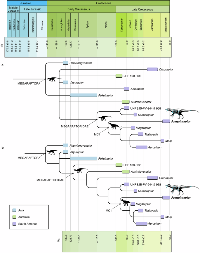
Phylogenetic analysis 1 included all operational taxonomic units within the matrix, whereas analysis 2 excluded the fragmentary taxon Aoniraptor. Both analyses postulated Megaraptora as well nested within Coelurosauria as the sister group of Tyrannosauroidea (Supplementary Note 10 and Supplementary Fig. 21). Analysis 1 (Fig. 3a) found 102 most parsimonious trees (MPTs) of 1477 steps, with a Consistency Index (CI) of 0.303 and a Retention Index (RI) of 0.594. In the strict consensus of these MPTs, the Thai taxon Phuwiangvenator is placed at the base of Megaraptora, followed by a polytomy that includes the Asian forms Vayuraptor and Fukuiraptor, Australovenator and the fragmentary specimen LRF 100–10616 from Australia, Orkoraptor and Aoniraptor from Argentine Patagonia, and a clade containing all remaining South American megaraptorans included in the analysis (=Megaraptoridae following the phylogenetic definition employed herein). Relationships among this latter clade are generally well resolved, albeit with low Bremer support values (=1). More specifically, the generically unidentified megaraptorid from the Bajo Barreal Formation (represented by specimens UNPSJB-PV 944 and UNPSJB-PV 958; see ref. 7) is recovered as the sister taxon of Murusraptor. Relative to those of other Patagonian Upper Cretaceous stratigraphic units, the dinosaur fauna of the Bajo Barreal Formation is frequently regarded as being comprised by comparatively plesiomorphic representatives of its constituent clades31,92; therefore, the early branching position of the Bajo Barreal taxon within Megaraptoridae is not unexpected. Both these Patagonian forms in turn constitute the sister taxon of a diverse clade of derived South American megaraptorids. This clade, herein informally termed Megaraptorid Clade 1 (=MC1) for ease of discussion, is united by the ambiguous synapomorphy of the parietals being narrow between the supratemporal fossae, with each reduced to about 10–30% the mediolateral width of the fossa (character 265, state 1; present in Joaquinraptor and Megaraptor). Within MC1, Joaquinraptor is placed as an early diverging form.
a Strict consensus of most parsimonious trees (MPTs) recovered by analysis 1, which included all taxa in the phylogenetic data matrix. b Strict consensus of MPTs recovered by analysis 2, which was conducted after pruning Aoniraptor libertatem. Note general geographic and stratigraphic congruence of taxa in (b), with earliest diverging megaraptorans known from the Early Cretaceous (Valanginian–Aptian) of Asia (Thailand and Japan), earliest diverging megaraptorids from the mid-Cretaceous (Cenomanian–Turonian) of Australia (New South Wales and Queensland), and derived megaraptorids from South America (Argentinean Patagonia), mostly from the Late Cretaceous (Coniacian–Maastrichtian). Abbreviation: MC1 Megaraptorid Clade 1. Silhouettes at selected nodes by T. Tischler (Megaraptora; https://www.phylopic.org/images/c6099e64-f64e-4685-89a8-10185713ba65/australovenator-wintonensis), Jagged Fang Designs (Megaraptoridae, https://www.phylopic.org/images/236a6e22-f960-46c2-acf1-3fc23c38a60a/aerosteon-riocoloradensis), and T. Dixon (MC1; https://www.phylopic.org/images/df11d20b-642f-4af2-b524-7a6c073a29b7/maip-macrothorax), respectively.
Analysis 2 (Fig. 3b), conducted after Aoniraptor was pruned, yielded only 12 MPTs of 1476 steps, with a CI of 0.307 and an RI of 0.602. The resulting strict consensus tree shows improved resolution, particularly at the base of Megaraptora. Phuwiangvenator is recovered in the same position as in analysis 1, but the abovementioned polytomy is largely resolved, leaving only Vayuraptor and Fukuiraptor as the next-latest diverging megaraptorans. The Australian forms LRF 100–106 and Australovenator are postulated as early diverging megaraptorids and successive sister taxa to a clade comprising all South American megaraptorids in the analysis. This latter group is united by the absence of mesial denticles (character 2, state 2). The recovery of this clade—consisting entirely of Argentinean megaraptorids reaching or exceeding approximately 7 m in total body length—supports the hypothesis that Megaraptoridae experienced a substantial increase in body size upon their arrival in South America during the mid-Cretaceous7 (see also below). The results of analysis 2 postulate Orkoraptor as the earliest diverging member of the Argentinean clade. As in analysis 1, the remaining South American megaraptorids are situated within a clade that is itself divided into two subclades, UNPSJB-PV 944 & 958 + Murusraptor and MC1. As in analysis 1, Joaquinraptor is recovered as the earliest diverging member of MC1.
In order to down-weight characters that exhibit widespread homoplasy, we applied extended implied weighting (k = 3) to both analyses. Similar results were obtained for analysis 1, but (after pruning Aoniraptor) analysis 2 yielded better resolution within Megaraptoridae (see Supplementary Fig. 21c). Among derived megaraptorids, both the clades UNPSJB-PV 944 & 958 + Murusraptor and MC1 were again recovered; however, there is improved resolution among derived members of MC1, with Maip being postulated as the sister taxon of Tratayenia rosalesi + Aerosteon.
In accordance with the study of Aranciaga Rolando et al.8, both our phylogenetic analyses (analysis 1 and analysis 2) recovered nested clades of South American megaraptorids, one more inclusive than the other. Here, we compare the results of our analysis 2 (i.e., our analysis that excluded Aoniraptor) to those of the analysis without Aoniraptor and the Bajo Barreal Formation megaraptorid (the latter represented by specimens UNPSJB-PV 944 and UNPSJB-PV 9587) presented by Aranciaga Rolando et al.8. The topologies of the strict consensus trees recovered by these two analyses differ with regard to the hypothesized placements of selected Patagonian megaraptorids. In our analysis, Megaraptor occupies a relatively derived position, whereas Orkoraptor is supported as an earlier diverging form. Therefore, instead of the clade Megaraptor + Murusraptor + “Clade B” recovered by Aranciaga Rolando et al.8 (i.e., these authors’ “Clade A”), we postulate Orkoraptor as the outgroup to an unnamed clade that itself includes the sister taxa UNPSJB-PV 944 & 958 + Murusraptor and MC1 (the latter of which encompasses all megaraptorid taxa within Aranciaga Rolando et al.’s8 “Clade B,” with the addition of Joaquinraptor and the aforementioned exception of Orkoraptor).
In sum, our phylogenetic analyses yielded several notable results that may be pertinent to future discussions of megaraptoran systematics. First, and unsurprisingly given the data matrix we used, the analyses support the placement of Megaraptora within Coelurosauria, as the sister group of Tyrannosauroidea. Moreover, Joaquinraptor is recovered as the earliest diverging member of a newly identified clade of derived South American megaraptorids, herein termed Megaraptorid Clade 1 (=MC1). Nevertheless, although the position of Joaquinraptor within Megaraptoridae on the whole is well supported, MC1 is presently supported by only a single synapomorphy; furthermore, two putative members of the clade (Maip and Tratayenia) are relatively incompletely preserved, being presently known only from postcranial (mainly postcranial axial skeletal) bones. As such, future analyses should evaluate the proposed monophyly and taxonomic content of MC1 and, therefore, its potential systematic validity, as well as the specific relationships of taxa within the clade.
Implications for Patagonian theropod faunal provincialism and megaraptorid palaeobiology
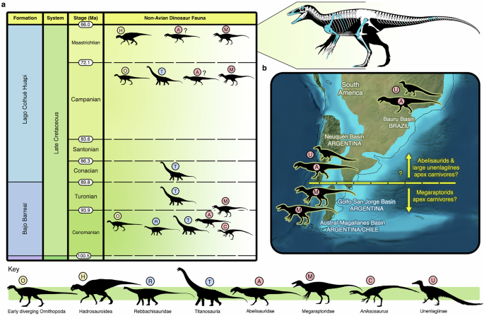
The post-Turonian central (Fig. 4a) and southern Patagonian fossil record of large-bodied non-avian theropod dinosaurs is, at the moment, numerically dominated by megaraptorids7,8,22,23,31,53. Nevertheless, to date, all described central and southern Patagonian megaraptorid specimens have been fragmentary; therefore, Joaquinraptor is important in being, by a substantial margin, the most complete representative of the clade yet found from this geographic area. Moreover, this taxon was recovered from only a few meters below the top of the Lago Colhué Huapi Formation, and is therefore Maastrichtian (probably late Maastrichtian) in age, rendering it the latest surviving megaraptoran yet known from central Patagonia and one of the stratigraphically youngest (quite possibly the youngest) members of the group found anywhere in the world. Although more evidence is needed, Patagonian megaraptorids present a suite of putative synapomorphies, such as the absence of mesial denticles from the tooth crowns, a manual ungual I that is subequal in length to the ulna, and a well-developed “mediolateral platform” on the flexor tubercle of this ungual8,35, that suggest that these theropods may have been evolutionarily and geographically differentiated from earlier diverging members of the group (e.g., Australovenator).
a Upper Cretaceous non-avian dinosaur faunas of the Chubut Group, Golfo San Jorge Basin, central Patagonia, Argentina, indicating stratigraphic position of Joaquinraptor casali gen. et sp. nov. (denoted by skeletal reconstruction). b Hypothesized distribution of medium- to large-bodied theropod dinosaurs occupying apex predator niches in selected South American depositional basins during the Campanian and Maastrichtian stages of the Late Cretaceous. Note absence of Megaraptoridae north of the Golfo San Jorge Basin. Palaeogeographic basemap ©2023 Colorado Plateau Geosystems Inc., used with permission. Dinosaurian silhouettes except Megaraptoridae by S. Hartman (early diverging Ornithopoda, https://www.phylopic.org/images/05b2229f-34dd-4fdc-aefb-8158a6b38119/parksosaurus-warreni; Hadrosauroidea, https://www.phylopic.org/images/4ae90b8f-bbb4-47cd-a914-98e1c845cf1e/gryposaurus-monumentensis; Rebbachisauridae, https://www.phylopic.org/images/a5b8df09-f5f9-43ae-ae2a-7822ff3e3d56/nigersaurus-taqueti; Titanosauria, https://www.phylopic.org/images/33ae8000-0046-41bb-b68a-189a0eed3eca/opisthocoelicaudia-skarzynskii; Abelisauridae, https://www.phylopic.org/images/d82ad39a-0770-4f85-99a6-f1d17bd1bf64/majungasaurus-crenatissimus), FunkMonk (Aniksosaurus, https://www.phylopic.org/images/d3ae8c0d-107d-45de-992e-177c551e079d/coelurus-agilis), and T. Dixon (Unenlagiinae, https://www.phylopic.org/images/eb481f28-4f1a-4cfc-96ea-f3ab0a622e6b/austroraptor-cabazai).
Furthermore, the discovery of Joaquinraptor supports the hypothesis that megaraptorids were the apex predators and the most abundant large-bodied theropods in the latest Cretaceous (Campanian–Maastrichtian) of central and southern Patagonia, contrasting with penecontemporaneous faunas from more northerly areas of South America that appear to have been dominated by abelisaurids and large unenlagiines (Fig. 4b; see also refs. 7,8,23,93). Novas et al.23 suggested that this putative theropod provincialism may have been related to palaeoenvironmental differences. Notably, however, the sedimentology of the middle (i.e., Santonian–?lower Maastrichtian) section of the Lago Colhué Huapi Formation, which has yielded several other megaraptorid fossils (31,55; Fig. 4a), is characterized by fluvial channel morphologies that indicate that the fauna it preserves lived inland, well away from the palaeoshoreline; moreover, clay analyses support a seasonally dry or semiarid climate for this part of the formation (see ref. 46 and references therein). Conversely, in the uppermost section of the formation, at a stratigraphic level close to that from which Joaquinraptor was collected, sedimentological and palynological inferences support the existence of a warm, humid floodplain palaeoenvironment close to the sea (see Supplementary Note 3). Consequently, it appears that, at least in central Patagonia, megaraptorids endured significant palaeoclimatic changes and successfully adapted to fluctuating palaeoenvironmental conditions through Late Cretaceous time, raising the possibility that these theropods were able to thrive in a broad range of habitats. As such, alongside putative palaeoenvironmental distinctions, additional factors, such as the potential palaeogeographic isolation of central and southern Patagonia from more northern areas of South America by epicontinental seas (Fig. 4b; see also, e.g., Reguero and Goin94 [these authors’ Fig. 8]) and/or pronounced floristic differences (see, for example, the reconstructed Late Cretaceous palynological bioprovinces proposed by Vajda and Bercovici95 [these authors’ Fig. 3b]) may also have driven the differentiation of large-bodied theropod faunas in southern South America at the end of the Mesozoic.
The distinctive megaraptorid forelimb may have played an important role in the apparent adaptability of these theropods, perhaps allowing them to exploit resources in varying ways and/or to feed on an array of vertebrate taxa. For example, megaraptorids may have used their hypertrophied manus with its enlarged, trenchant unguals to access soft tissues96 and/or to aid in prey capture and manipulation9,20,25,97,98. In addition, several features of the megaraptorid antebrachium, such as the proximodistally elongate olecranon process (the insertion area of M. triceps brachii) and the vertical ridge on the proximolateral surface of the ulna (a probable osteological correlate for the M. anconeus insertion9,99,100), may have conferred enhanced extension and flexion capabilities.
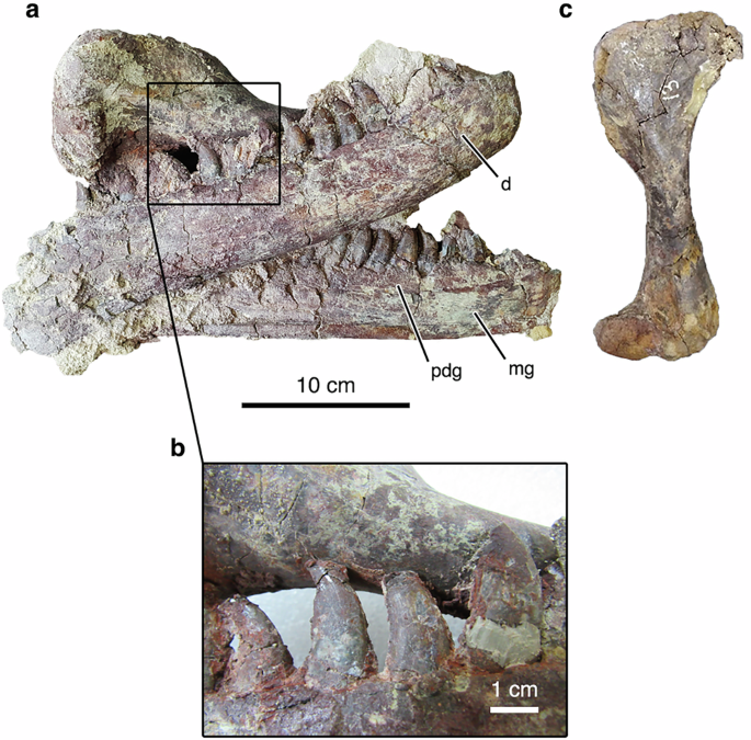
As mentioned above, in addition to the Joaquinraptor skeleton, a crocodyliform right humerus was recovered from the type quarry of the new megaraptorid. Interestingly, this bone was situated between the closely associated left and right dentaries of the Joaquinraptor holotype, with the apices of several tooth crowns of the megaraptorid in direct contact with the humeral shaft, which also shows potential tooth marks (Fig. 5); moreover, this humerus is the only non-megaraptorid vertebrate fossil yet identified from the site. As such, this discovery may constitute direct evidence regarding prey selection within Megaraptoridae (although isolated megaraptorid teeth have been found in association with other saurischian dinosaur specimens, potentially also indicating feeding behavior [e.g., refs. 41,101]). Nevertheless, this megaraptorid/crocodyliform co-occurrence could conceivably be instead related to other behaviors, such as interspecific combat or competition for food, or even just a coincidental association (e.g., the humerus was moved to a position between the Joaquinraptor dentaries by water currents). Detailed taphonomic studies are currently underway to assess the probable cause and potential significance of this association.
a Dentaries of Joaquinraptor (UNPSJB-PV 1112) and an associated crocodyliform right humerus (UNPSJB-PV 1113) in the position in which they were recovered prior to their mechanical separation in the laboratory (right dentary in lateral view, left dentary in medial view, humerus in posterior view [proximal end only]). b Detail of a showing a portion of the crocodyliform humeral shaft (UNPSJB-PV 1113) in contact with the right dentary tooth crowns of Joaquinraptor (UNPSJB-PV 1112). c Crocodyliform right humerus (UNPSJB-PV 1113) in anterior view. Abbreviations: d dentary, mg Meckelian groove, pdg paradental groove.
In sum, the singular morphology of the megaraptorid forelimb may have played an important role in the evolutionary success of these theropods in the Late Cretaceous of central and southern Patagonia. The specialized development and capabilities of the forelimb were almost certainly adaptations related to prey capture9,97,98, and, in the case of Joaquinraptor, these prey items may have at least occasionally included crocodyliforms (Fig. 5).
Megaraptorid palaeobiogeography and body size evolution
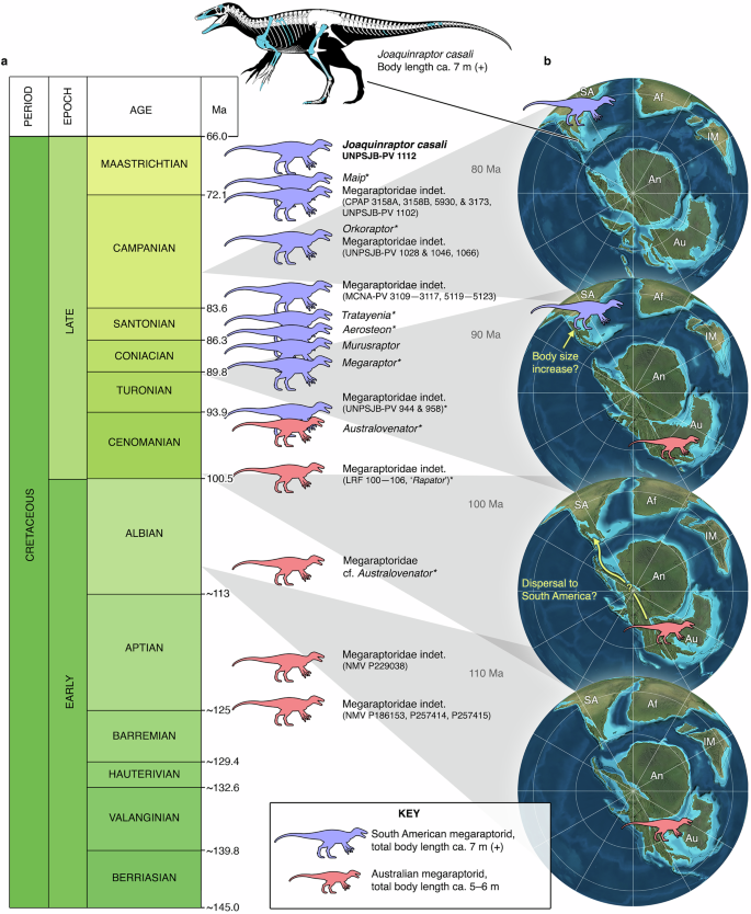
Based on the fossil record of Megaraptoridae as it is currently understood, Lamanna et al.7 hypothesized that South American members of the clade underwent a substantial increase in body size with respect to their earlier diverging and mostly stratigraphically older Australian relatives. Specifically, megaraptorids are proposed to have experienced an increase in size from approximately 5 m in total body length and 300 kg in body mass to more than 7 m in length and 1000 kg in mass not long after their dispersal to South America from Australia, presumably via Antarctica, during the mid-Cretaceous7. Moreover, South American megaraptorids are hypothesized to have maintained this elevated body size until their extinction at or near the end of the Cretaceous7,8.
The nearly complete (missing only the medial condyle of the distal end) left femur of the holotype of the Maastrichtian megaraptorid Joaquinraptor has a total proximodistal length of 685 mm and a circumference (measured immediately distal to the fourth trochanter) of 275 mm (Table 2). Based on the scaling equation presented by Campione and Evans102 and employed in many subsequent works (e.g., refs. 7,103), this circumference yields a mass estimate of 1036 kg for this individual. Approximately the same circumference (280 mm) was obtained from the fragmentary femur of another large-bodied megaraptorid specimen from a nearby exposure of the stratigraphically older (Cenomanian–Turonian) Bajo Barreal Formation (UNPSJB-PV 958), from which an estimated body mass of 1089 kg was derived7. Furthermore, a mass estimate that is precisely the same as that generated for Joaquinraptor (1036 kg) was proposed for MUCPv 1353, an undescribed partial megaraptorid skeleton (probably referable to Megaraptor) from the Turonian–Coniacian Portezuelo Formation of northwestern Patagonia7. On the other hand, the type specimen of the early diverging Australian megaraptorid Australovenator has a femur length of 578 mm15, whereas that of the holotype of the non-megaraptorid megaraptoran Fukuiraptor is 507 mm2. Likewise, the estimated body mass of the former taxon (314 kg103) is approximately 3.3–3.5 times less than that of Joaquinraptor, MUCPv 1353, and UNPSJB-PV 9587. Aranciaga Rolando et al.8 proposed a body length of more than 9 m for Maip, a megaraptorid from the Maastrichtian Chorrillo Formation of southern Patagonia, which would likely establish this taxon as the largest-bodied megaraptorid yet known.
In sum, Joaquinraptor accords with Lamanna et al.’s7 hypothesis of body size evolution in South American megaraptorids in that it was probably longer than 7 m and heavier than 1000 kg; in other words, approximately the same size as MUCPv 1353 and UNPSJB-PV 958. Furthermore, Joaquinraptor is from the Maastrichtian—probably the late Maastrichtian—supporting the hypothesis that Megaraptoridae maintained this elevated body size until their extinction (Fig. 6).
a Stratigraphic distribution of definitive megaraptorid fossils through the Cretaceous. Asterisks adjacent to some records indicate that additional, generically indeterminate megaraptorid materials have been reported from the same stratigraphic unit. Stratigraphic position of Aerosteon riocoloradensis within the upper Coniacian–lower Campanian is uncertain (see ref. 21). b Hypothesis of palaeobiogeography and body size evolution in Megaraptoridae. Megaraptorids are considered to have originated in eastern Gondwana (Australia) during the Early Cretaceous, then dispersed to western Gondwana (South America) via Antarctica during the mid- (i.e., late Early or early Late) Cretaceous (see refs. 7,8,16). Red silhouette represents Australian megaraptorid ca. 5–6 m in total body length; blue silhouette represents ca. 7 m (+) South American megaraptorid. Cretaceous timescale in a from ref. 110; palaeogeographic reconstructions in b are south polar views at (bottom to top) 110, 100, 90, and 80 Ma (©2023 Colorado Plateau Geosystems Inc., used with permission).
Recently, Kotevski et al.19 proposed that selected megaraptorid postcranial elements (a set of two articulated caudal vertebrae and their corresponding haemal arches [NMV P257414], an isolated partial manual ungual II [NMV P186153], and the distal end of a tibia [NMV P257415]) from the upper Barremian–lower Aptian upper Strzelecki Group of Victoria, Australia suggest that ca. 6 m individuals of this theropod clade inhabited that continent during the Early Cretaceous. The upper Strzelecki Group bone that implies the largest individual—a megaraptorid perhaps slightly greater than 6 m in body length—is the fragmentary manual ungual II NMV P186153, which was also described and/or analysed at length by Benson et al.104, Poropat et al.17, and White and Campione105. That said, this claw is isolated and incomplete, and as such, the hypothesized body size of the individual to which it pertained—though it may well be correct—cannot be considered to be robustly documented given the fragmentary nature of the currently available evidence. Consequently, we consider the hypothesis that >6 m megaraptorids were present in Australia to be only tentatively supported at present. Moreover, even if NMV P186153 does indeed represent a megaraptorid marginally greater than 6 m in body length, the likelihood remains that all definitive South American megaraptorid records indicate animals that are still larger (ca. 7 m [+] in length and ca. 1000 kg [+] in mass). As such, in our view, Lamanna et al.’s7 hypothesis that megaraptorids experienced a substantial increase in body size shortly after their arrival in South America remains consistent with the presently available data (Fig. 6). That said, it may potentially be only a matter of time before definitive evidence of large-bodied (≥7 m, ≥1000 kg) megaraptorids is discovered in the Early or mid-Cretaceous of Australia.
To conclude, as proposed by Lamanna et al.7 and supported by more recent discoveries (e.g.,8,106), including Joaquinraptor (Fig. 6), South American megaraptorids appear to have attained larger body sizes (~7 m or greater in total body length) than their generally more ancient relatives from Australia (~5–6 m19). Patagonian megaraptorids may have continued to increase in size through the remainder of their evolutionary history—perhaps spurred at least in part by the extinction of carcharodontosaurids during the Turonian8,106—with some latest Cretaceous forms (e.g., Maip) potentially reaching body lengths of 9 m or more8. Among other benefits, this may have allowed megaraptorids to occupy the apex predator niches in their respective habitats. Moreover, and as evidenced by the Maastrichtian (probably late Maastrichtian) stratigraphic occurrence of Joaquinraptor, these theropods may well have survived to the Cretaceous/Palaeogene boundary, only to ultimately perish alongside all other non-avian dinosaurs.
Source link
