Researchers uncovered blood vessels in Scotty, the largest T. rex fossil. High-powered x-rays revealed mineralized tissues that deepen our understanding of dinosaur biology.
Although much of modern paleontology is devoted to searching for organic traces in fossils, no sample of dinosaur DNA has ever been recovered.
Most of what scientists know about dinosaurs comes from their bones and teeth preserved in the rock record. While these hard tissues are invaluable, they offer only limited insight into the biology and appearance of the animals themselves.
By contrast, soft tissues are exceedingly rare in fossils but can provide a much richer and more detailed picture of ancient life. Examples include preserved muscles, ligaments, pigments, or even skin impressions such as scales or feathers, which reveal information about how dinosaurs lived and what they looked like.
Another interesting soft tissue that can be found in bones are blood vessels. My research team and I discovered blood vessels preserved in a Tyrannosaurus rex fossil, and our findings were recently published in Scientific Reports.
As an undergraduate physics student at the University of Regina, I joined a research team using particle accelerators to study fossils. There, I first discovered blood vessels in a bone from a T. rex using advanced 3D models. It’s been nearly six years since that moment; I am now working on my PhD where I use my background in physics to advance analysis techniques in fossil research.
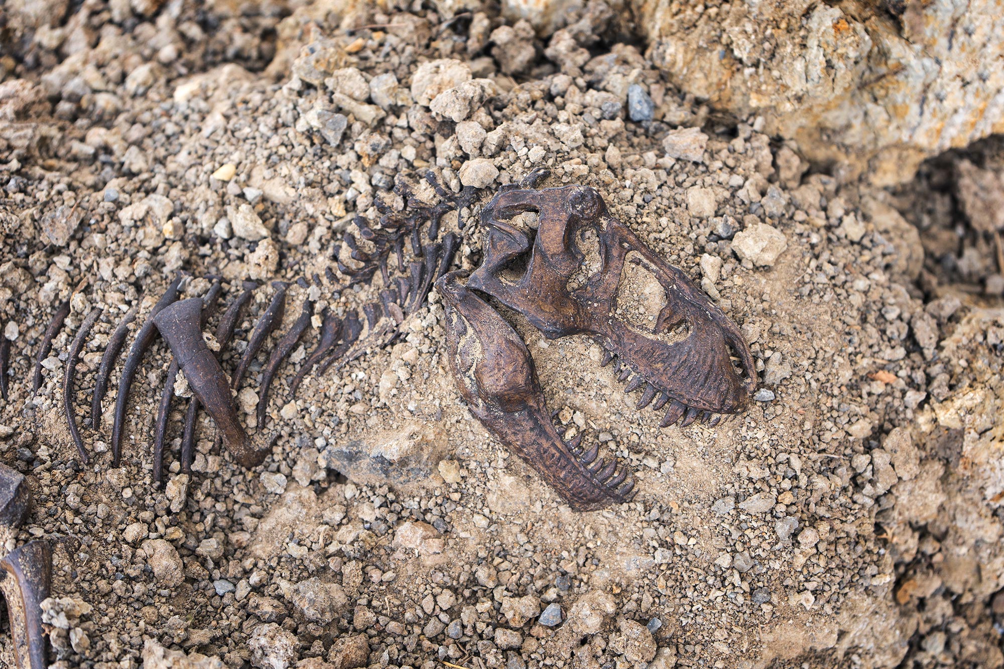
An extraordinary specimen
The preserved vessels came from an exceptional T. rex specimen nicknamed Scotty. Housed in the Royal Saskatchewan Museum in Canada, Scotty is the largest T. rex ever discovered and remains one of the most complete skeletons of the species.
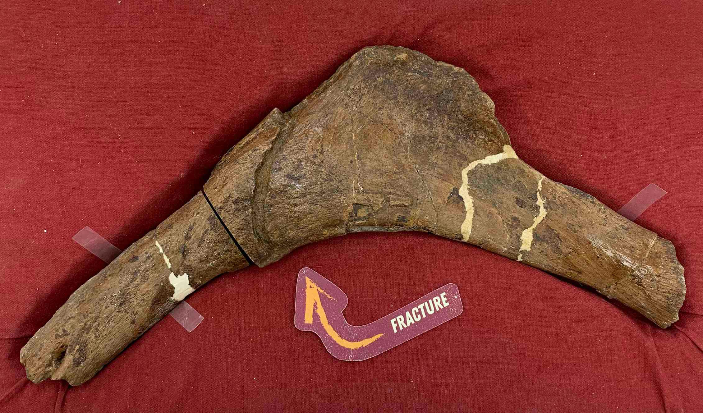
Evidence from the bones suggests Scotty lived a rough life 66 million years ago, with many injuries that may have resulted from combat with another dinosaur or from illness. One rib in particular displayed a large fracture that had partially healed before the animal’s death.
In general, after bones experience a traumatic event like a fracture, there is a huge increase in the activity of blood vessels in the affected area as part of the healing process. We believe this is what was found in Scotty’s rib: an extensive network of mineralized vessels that we were able to examine using reconstructed 3D models.
Revolutionizing paleontology research
When analyzing fossil bones, there are two main challenges. The first is how to examine the interior of the bones without damaging the fossil. And second, the bones are very large and can be quite dense due to the fossilization process, where minerals replace and fill in original organic materials.
At first, we thought we could perform an computed topography (CT) scan of the bone, similar to what is used for medical purposes, which allows imaging of bones without damaging them. While this solves the first problem, the second problem means that a conventional medical CT machine is not nearly powerful enough to penetrate the dense bone.
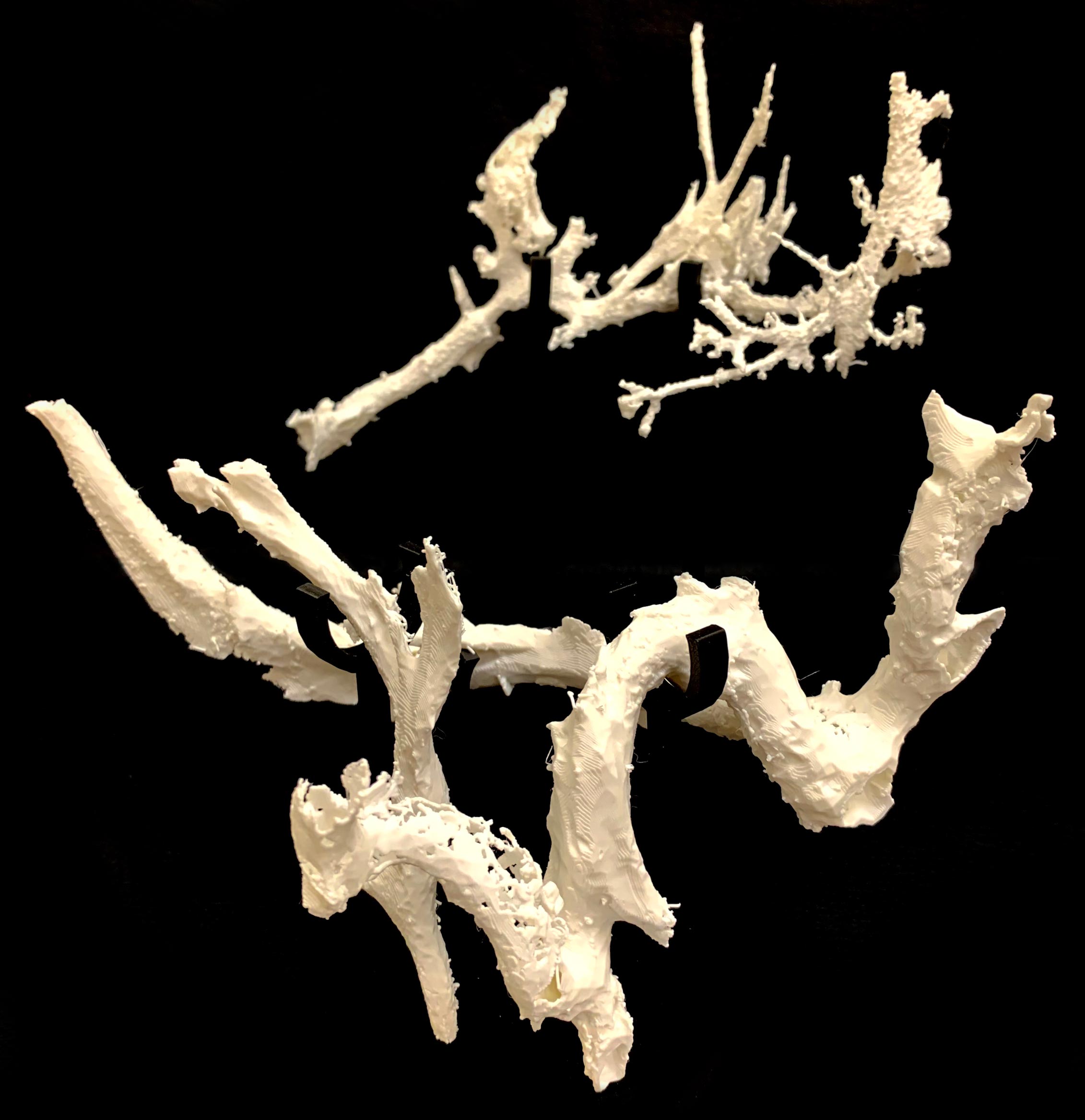
For our examination, we used synchrotron light, special high-intensity x-rays. These are produced at select particle accelerator labs, and allow us to investigate microstructures such as blood vessels in the bone with ease.
Synchrotron x-rays can also be useful for chemical analysis. We found the vessels were preserved as iron-rich mineralized casts, a common form of fossilization, but in two distinct layers. This layering is due to the complicated environmental history that led to the exceptional preservation seen in Scotty’s rib.
Written in blood vessels
By analyzing blood vessels produced by an incompletely healed fracture, we can hopefully learn how T. rex healed, helping speculation on how Scotty was able to survive after sustaining injuries. This could lead to evolutionary information comparing the vessel structures seen in Scotty to other dinosaur species, as well as modern relatives to dinosaurs like birds.
The results may also help future fossil exploration by guiding scientists to target bones that show signs of injury or disease, potentially increasing the chances of discovering more vessels or other types of preserved soft tissues.
With cross-disciplinary research and novel applications of advanced technologies, there is so much potential to recreate the past lives of dinosaurs like never before.
Reference: “In situ analysis of vascular structures in fractured Tyrannosaurus rex rib” by Jerit L. Mitchell, Mauricio Barbi, Ryan C. McKellar, Monica Cliveti and Ian M. Coulson, 4 July 2025, Scientific Reports.
DOI: 10.1038/s41598-025-06981-z
Adapted from an article originally published in The Conversation.![]()
Jerit Leo Mitchell receives funding from Mitacs Accelerate and the Sylvia Fedoruk Centre for Nuclear Innovation.
Never miss a breakthrough: Join the SciTechDaily newsletter.
Source link


