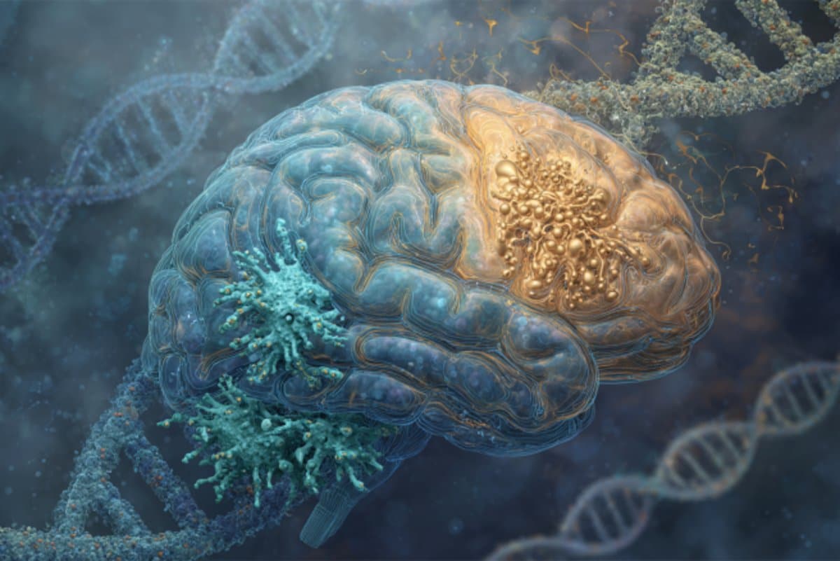
Summary: Researchers analyzing post-mortem brain tissue found two cell types altered in people with depression: excitatory neurons that regulate mood and stress, and microglia that manage inflammation. The findings highlight how depression is tied to measurable brain changes, not just emotional symptoms.
By mapping gene activity alongside DNA regulation, scientists identified key cellular disruptions unique to depression. This breakthrough could lead to therapies that target specific cells, offering hope for millions affected worldwide.
Key Facts
- Targeted Cell Types: Mood-related excitatory neurons and inflammation-regulating microglia show altered gene activity in depression.
- Rare Resource: The study used brain samples from the Douglas-Bell Canada Brain Bank, one of few with psychiatric condition donations.
- Therapeutic Potential: Identifying these cells opens a pathway to precision treatments for depression.
Source: McGill University
Researchers at McGill University and the Douglas Institute have identified two specific types of brain cells that are altered in people with depression.
The study, published in Nature Genetics, opens the door to developing new treatments that target these cells and deepens our understanding of depression, a leading cause of disability worldwide that affects more than 264 million people.

“This is the first time we’ve been able to identify what specific brain cell types are affected in depression by mapping gene activity together with mechanisms that regulate the DNA code,” said senior author Dr. Gustavo Turecki, a professor at McGill, clinician-scientist at the Douglas Institute and Canada Research Chair in Major Depressive Disorder and Suicide.
“It gives us a much clearer picture of where disruptions are happening, and which cells are involved.”
Rare brain bank enables breakthrough
The researchers used post-mortem brain tissue from the Douglas-Bell Canada Brain Bank, one of the few collections in the world with donated tissue from people who had psychiatric conditions.
They used single-cell genomic techniques to analyze RNA and DNA from thousands of brain cells, identifying which cells worked differently in depression and what DNA sequences could explain those differences. They studied samples from 59 people who had depression and 41 people without it.
The results revealed altered gene activity in a certain type of excitatory neuron involved in mood and stress regulation, and in a subtype of microglia cells, which help manage inflammation. In both cell types, many genes were functioning differently in people with depression, suggesting potential disruptions in these key brain systems.
By pinpointing brain cells affected in depression, the study adds new insight into its biological basis and, more broadly, challenges lingering misconceptions about the disorder.
“This research reinforces what neuroscience has been telling us for years,” Turecki said. “Depression isn’t just emotional, it reflects real, measurable changes in the brain.”
As a next step, the researchers plan to study how these cellular changes affect brain function and whether targeting them could lead to better therapies.
About the study
Funding: The study was funded by Canadian Institutes of Health Research, Brain Canada Foundation, Fonds de recherche du Québec – Santé and Healthy Brains, Healthy Lives initiative at McGill University.
About this neuroscience and depression research news
Author: Keila DePape
Source: McGill University
Contact: Keila DePape – McGill University
Image: The image is credited to Neuroscience News
Original Research: Closed access.
“Single-nucleus chromatin accessibility profiling identifies cell types and functional variants contributing to major depression” by Anjali Chawla and Gustavo Turecki et al. Nature Genetics.
Abstract
Single-nucleus chromatin accessibility profiling identifies cell types and functional variants contributing to major depression
Genetic variants associated with major depressive disorder (MDD) are enriched in the regulatory genome.
Here, we investigate gene-regulatory mechanisms underlying MDD compared to neurotypical controls by combining single-cell chromatin accessibility with gene expression in over 200,000 cells from the dorsolateral prefrontal cortex of 84 individuals.
MDD-associated alterations in chromatin accessibility were prominent in deep-layer excitatory neurons characterized by transcription factor (TF) motif accessibility and binding of NR4A2, an activity-dependent TF reactive to stress.
The same neurons were enriched for MDD-associated genetic variants, disrupting TF binding sites linked to genes that likely affect synaptic communication.
Furthermore, a gray matter microglia cluster exhibited decreased accessibility in individuals with MDD at binding sites bound by TFs known to regulate immune homeostasis.
Finally, we identified gene-regulatory effects of MDD-risk variants using sequence-based accessibility predictions, donor-specific genotypes and cell-based assays.
These findings shed light on the cell types and regulatory mechanisms through which genetic variation may increase the risk of MDD.
Source link

